What is the Best Choice of Whitening Lamp for Dental Practice

Constant Temperature Whitening Lamp

How to Select the Best Type of Whitening Lamp for Your Practice
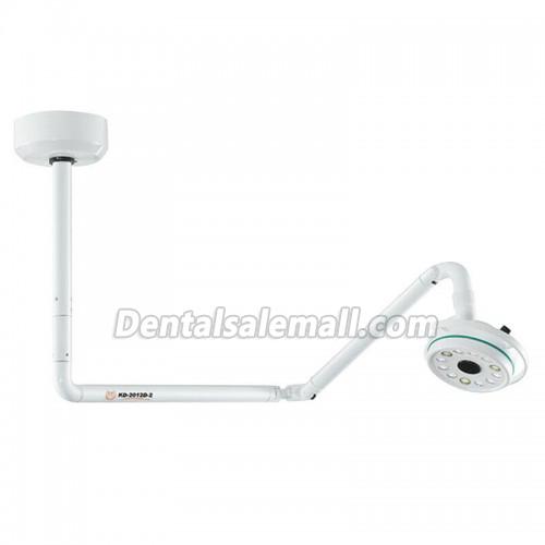
The best type of dental lamp for your practice should be chosen based on the specific needs and preferences of the dentist.
There are many different types of lamps that can be used in a dental office. They vary in terms of light output, color temperature, and size.

5 Tips on How to Care for a Dental Lamp
1) The most important thing to remember is to keep the lamp clean. When you wipe it down, be sure to use a damp cloth and not a dry one. If you use a dry cloth, you risk scratching the surface of the lamp and that can lead to dirt particles getting into the light fixture.
2) Make sure that you change your lamp bulb at least once a year. This is especially true if you have an older style of dental appliance light.
3) If your dentist recommends it, make sure that you get your lamp professionally cleaned every three months or so. This will help ensure that it stays in good shape for years to come and doesn’t need replacement too soon.
4) Be careful with how much water you put on the top of the dental lamp when cleaning

3 Common Problems You May Encounter with a Dental Lamp
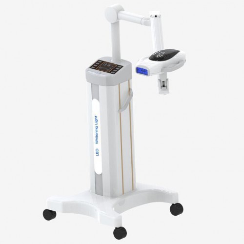
A mobile dental whitening lamp is a type of light that is used in dentistry. It is used to provide a bright, white light that can be focused onto an area. Dental lamps are often used in conjunction with dental tools like drills and picks.
Just like any other device, a dental lamp may have problems from time to time. This article will explore three common problems you may encounter with a dental lamp and what to do about them!


Why Trust Your Practice Success to an Oral Health Light Source?
Oral health light sources are a new technology that is changing the way we see oral health. This technology has been around for a while, but it is now being used as a replacement to traditional dental lights. The benefits of this new technology are many and include:
-A more comfortable experience for patients
-Reduced dental costs
-The ability to treat patients in areas without electricity or water
-The ability to take x-rays with less radiation.

The Difference Between a Dental PMMA & PEAK Block

What is a Dental PMMA Block and Why Should I Use It?
Dental PMMA Blocks are a material that is becoming increasingly popular in the dental industry. They are used by dental labs to replace amalgam fillings.
Dental pmma blocks are made from a resin that has been mixed with an aluminum oxide. This combination creates a strong, but flexible material that is easy to shape and work with. It also means that it can be easily molded into different shapes and forms without losing its strength.
The use of dental pmma blocks is one of the most important developments in dentistry today as they can provide patients with more options for treatment as well as reduce the amount of anesthesia needed during treatment procedures.

PEAK Block is a type of dental block that has been specially designed for use with prosthodontics. It was created by Dr. Michael Pfeifer, who wanted to improve the quality of his patients’ dental care by making it more comfortable and easier to perform procedures.

What are the Differences between a PMMA and PEAK Block?
A PMMA or a PEAK block is a type of dental crown that is used to cover the entire tooth and restore its shape. However, they have different uses and purposes.
PMMA blocks are used in cases where you want to create a new tooth structure while dental peak blocks are used in cases where you want to restore an existing tooth structure.

The use of a dental P-MMA block is a great way to reduce tooth extraction times in emergency dental cases like tooth loss. This type of block has been shown to reduce the need for anesthesia, sedation and pain medications. It also improves patient outcomes and reduces cost for the dentist.

Advantages of using a dental P-MMA block:
1) Reduces need for anesthesia, sedation, pain medications 2) Improves patient outcomes 3) Reduces cost for the dentist.

6 Reasons to Start Using a Dental PMMA Block Today
The dental cad cam block is a type of dental material that is used to make impressions of teeth and create molds. It is made of a silicone rubber that is hard, durable, and provides a smooth surface for the impression.
Some of the benefits of using this dental lab block are:

  • It makes it easier to work with the patient and provide them with more comfortable treatment;
  • It keeps teeth from becoming too sensitive or damaged during treatment;
  • This dental material can be easily removed from the patient’s mouth after treatment is done;

What should take into consider when choosing dental surgical Light

What is a Dental Surgical Light?
The dental surgical light is a tool that helps the dentist to get a better view of the oral cavity during procedures.

The dental surgical light is an essential tool for dentists, but the 3 main types of lights in dentistry today are wall mounted dental light, ceiling mounted dental light, post-mounted dental light and chair mounted dental light. You can choose one according to your own requirement.

How to Choose the Right Surgical Light for Your Practice
Choosing the right surgical light is an important aspect of any operating room. It helps to improve patient safety and reduce surgical time.
Surgical lights are typically LED lights that remain cool to the touch during use and are typically used for ophthalmic or general surgery. Some of them can also be used for dental procedures.
There are 3 main components that make up a surgical light: the power source, the lens, and the reflector. The power source can be either a corded or cordless battery, while the lens is either a metal halide or xenon lamp. The reflector is typically made of aluminum with a metal backing that reflects light back into the lamp.

Why Do You Need a High-quality Oral Light?
If you are a dentist, it is important that you have the best dentists’ lights. You will need them to make sure that your patients are comfortable and not in pain during their visit.
Dentists can use these lights for many different purposes:

  • Dental surgery procedures, such as cleaning and polishing teeth
  • Using the light to help with night vision
  • Helping patients see better during general checkups

What Are the Key Features of the Excellent Dentist Operatory Lights?
Dentist’s lights are essential for every dental practice. They provide a safe and comfortable environment to work in.
There are a variety of features that make the best dentist’s lights. Here is a list of some of the features that you should look for when buying dentists lights:
Safety – The best dentist’s lights should be safe for the patients and staff members. These lights should have low voltage and high wattage.
Durability – Dentist’s lights must be durable to last for long periods of time without any damage or defects. The light source must also be able to withstand high temperatures and humidity levels in order to work efficiently throughout the day.
Lighting – Dentist’s lamps need to provide sufficient lighting that is adjustable, providing adequate illumination on all surfaces in the room.

Some Information about Dental Implant Surface

Dental root form implants are manufactured from a highgrade titanium alloy, the surface of which consists of a micro layer of titanium oxide. The implant surface can also be treated by plasma spraying, acid etching, sandblasting or coated with HA. The removal of plaque and calculus deposits from these implant surfaces with Dental Instruments designed originally for cleaning natural tooth surfaces can result in major alterations to the delicate titanium oxide layer. Altering the surface topography by roughening the surface may enhance calculus and bacterial plaque accumulation.

Resulting scratches, cuts or gouges may also reduce the corrosion resistancy of titanium, and corrosion and mechanical debris can accumulate in the surrounding tissue. The aim of procedures for debriding dental implants should be to remove microbial and other soft deposits, without altering the implant surface, and thereby adversely affect biocompatibility. Increased surface roughness can lead to an increase in bacterial accumulation and resultant soft tissue inflammation. Because of the critical nature of the implant/soft tissue relationship, metal ultrasonic scaler tips, hand scalers or curettes should not be used as they have been shown to significantly alter the titanium surface.

Current methods for professional cleaning of implant or titanium transmucosal elements include the set of plastic ultrasonic tips( ultrasonic scaler ) or hand instruments followed by the prophy cup polishing method or various types of floss and buffing strips. The design of the permanently cemented super structure often does not allow adequate access for the prophy cup, especially in interproximal areas, and plastic instruments are not very efficient for the removal of plaque or mineralized deposits. In addition, the prophy cup and paste method may leave residual paste at the implant/soft tissue interface area.

Airpolishing consists of directing, water, air and sodium bicarbonate towards the tooth or implant surface, resulting in efficient removal of bacterial plaque and soft mineralized deposits. The residual powder is biocompatible and being soluble is not retained at the implant/soft tissue interface( dental implant machine ).

Two airpolishing systems are currently available. One system, typically available on the Dentsply Prophyjet? and Cavijet,? the EMS Airflow, and the Satelec units, delivers the air and powder, typically at 60-80 psi pressure through one nozzel and the water through a separate concentric nozzel. Some mixing of the streams takes place at the interface of the streams, but the centre of the stream consists essentially of dry powder. This “Biphasic” stream is directed at the tooth or implant surface. Several studies have investigated this system, and its effects on implant surfaces, and conclude that this system can result in significant changes to the implant surface.

The Information about Dental Air Cleaning

The air in dental surgeries has a variety of microbiological particulates and aerosols generated from ultrasonic scaler and high-speed drills. They vary in size from 0.5 to 5 microns in diameter and can remain airborne for many hours.

Dentists and their staff can easily inhale the viruses and bacteria contained within the aerosols, with facemasks offering no protection against this fine particulate pollution. Capturing these microorganisms reduces the risk of cross-infection – for the patient, the dentist and the team.

Dental Surgeries use chemical disinfectants to decontaminate hands, surfaces and instruments. While eliminating viruses, germs and fungal spores, disinfectants often contain toxic agents such as aldehydes (formaldehyde and glutaraldehyde) or phenol. Continuous low-level exposure to aldehydes can have negative health effects, such as breathing difficulties, memory impairment, eye and skin irritation and irregular heartbeat. Toxic compounds such as isopropanol, ethanol and n-propanol can also cause irritation of the respiratory tract and the mucous membranes.

Mercury Vapours

Recent research studies have found that both dentists and their staff have a higher than average level of mercury in their body. Mercury is used in the amalgam for routine dental fillings. Mercury transforms from a solid to a gas at room temperature. The gas (which is the most easily absorbed type of mercury) can be inhaled when amalgam is placed in the mouth or removed. Mercury is highly toxic and humans should not be exposed to it.

With this news and patients becoming more health conscious, requests for amalgam removals by dental amalgamator are rising steadily. It is therefore now more important than ever, for dentists to protect themselves and their team from this harmful substance.

Dental Air Conditioning

It is now commonplace for dentists to have air-conditioning systems installed. These installation systems are, however, often a source of contamination themselves, either because they are equipped with less then adequate filtration or because they are drawing in polluted air from outside without filtering it sufficiently. Indoor air contamination can be many times greater than external conditions, and dental air cleaning is required.

Prompted by an ever growing number of dentist offices as customers, Commercial Air Filtration specifies the IQAir Dental Series which has been developed to provide a flexible, cost effective, silent and low maintenance air cleaning solution for dental practices.

How to Choose the Best Dental X-ray Machine

Dental X-rays are one of the most important part of your regular dental treatment. Your dentist uses the specialized imaging technology to look for hidden tooth decay – also called cavities – and can show dental issues such as abscessed teeth, dental tumors, and cysts. The purpose of these machines is to see things that are not visible by visual examination of the mouth alone. Dentists can use the images produced to see the teeth as well as the bones and soft tissues around them. Finding cavities, examining teeth roots, viewing tooth development, and checking the underlying bone health are all functions performed by various dental x ray machines.

When contemplating the change to digital dental in your practice, the choices can be confusing for the dentist. Dental radiography has evolved from film and chemical developers into a highly technical process that involves various types of digital x-ray machines, as well as powerful dental software programs to assist the dentist with image acquisition and diagnostic analysis of the acquired images. When making the decision to purchase x-ray equipment, the doctor needs to research the available options thoroughly, in order to make an informed choice for the “right” machine for his or her practice.

The first question that a doctor should ask themselves is, “What is the main type of treatment that I provide my patients?” If you are a general practitioner, a standard 2D panorex will provide all of the imaging requirements needed for such treatments as caries detection, diagnosis of TMJ issues, OPG images, and images of the patients entire detention in a single x-ray. Many of the newer 2D panoramic units also offer extraoral bitewing imaging capability, which allows the dentist to obtain a bitewing image without putting a sensor or periapical film inside of the patient’s mouth.

The orthodontist requires a way to obtain the size and form of craniofacial structures in the patient. For this reason, a cephalometric extension on the imaging x-ray device is necessary to acquire images that evaluate the five components of the face, the cranium and cranial base, the skeletal maxillae, the skeletal mandible, and maxillary dentition. The cephalometric attachment offers images such as frontal AP and lateral cephs.

If the practice is concentrated in endodontic and implant treatment, then a CBCT machine is the most practical method of providing the doctor with diagnostic tools such as mandibular canal location, surgical guides, and pre-surgical treatment planning with the assistance of powerful 3D dental software applications. The patient is benefited by the reduced radiation exposure provided by these machines.

The Development of Root Canal Treatment

Root canal treatment by root canal treatment equipment is the process of going inside the pulp space and removing the infected, dead tissue. The procedure involves removing the damaged area of the tooth (the pulp), cleaning and disinfecting it and then filling and sealing it. The common causes affecting the pulp are a cracked tooth, a deep cavity, repeated dental treatment to the tooth or trauma.  The space is then disinfected and sealed with special materials.

Generally speaking, whatever the cause of root canal or pulpal disease, root canal or endodontic treatment will be necessary to save the tooth. All dentists receive training in endodontic treatment and can perform root canal procedures, but often a general dentist will refer individuals who need endodontic treatment to an endodontist, a root canal specialist.

Endodontists are dentists who have completed an additional two or more years of advanced residency training in the diagnosis and management of diseases and disorders of the dental pulp tester, and in the diagnosis of dental pain; their focus is therefore on saving teeth. In order to make a proper assessment and accurate diagnosis of which tooth is affected and exactly what is causing the pain, a thorough history and examination is necessary, together with a radiographic picture (x-ray) of the tooth or area.

Your dentist or endodontist will check your medical history and current medications to ensure your health and treatment safety. If you are very nervous, an oral sedative or anti-anxiety medication may be helpful — discuss the options with your dentist or endodontist ahead of time.

Preliminary treatment to remove the decay and the source of infection of the pulp is necessary, along with a determination of whether the lost tooth structure can be restored. If a fracture of the tooth has reached the pulp, or infection is associated with gum disease, it could be more difficult, if not impossible, to save the tooth.

Nowadays, root canal treatments are performed with advanced techniques and materials, making them far more comfortable and faster. After root canal treatment is complete, your restorative dentist will usually place a crown on your tooth to safeguard against fracture.


Some Information about Dental Amalgam Separator

Dental offices that place or remove amalgam fillings are required to install and properly maintain an amalgam separator. Depending on the brand, a separator can be purchased from virtually any supply vendor or purchased directly from the manufacturer. Whatever separator is purchased; it is important that the system is promptly installed in order to comply with the new regulations. Proper documentation management is an integral part of this program to ensure that a certificate of recycling is kept on file, and a replacement canister is purchased once the marked fill line is reached or 12 months from the date of installation, whichever occurs first.

The regulations do not specify a minimum amount of time needed before replacing a used filter/canister, but the regulations do state and mandate that the manufacturer guidelines for replacement be followed. Since each amalgam separator is required to conduct testing for the ISO certification based on a 12-month replacement maximum, most amalgam separators are required to be replaced every 12 months or once the canister is full. This not only ensures that the separator is functioning as certified, but also to prevent the separator from moving into bypass mode, which would allow the wastewater to flow unrestricted or filtered directly into the separator. The EPA recommends that an amalgam separator should be monitored monthly to ensure the canister is replaced per the manufacturer’s instructions for use, and that a backup canister is kept on site to ensure proper replacement is conducted at the appropriate time.

Most separators are compatible with both large- and small- capacity dental offices and can be used with dry vacuum or wet vacuum systems. That being said, it is best to check with the manufacturer or distributor to make sure the right amalgam separator system is purchased.

The amalgam separator is installed before the main vacuum line intersects with the plumbing in other parts of the building, and separates solids before reaching the wastewater. The typical plumbing configuration in a dental office involves a chairside trap for each chair and a central vacuum pump with a vacuum pump filter. Chairside traps and vacuum pump filters remove approximately 78% of dental amalgamator particles from the waste stream. These chairside traps cannot be cleaned or washed; they must be recycled to ensure that amalgam particles are properly managed.

Most separator designs rely on the force of the dental facility’s vacuum to draw wastewater into the separator. These separators are estimated to reduce the discharge of metals to POTWs by at least 8.8 tons per year, about half of which is comprised of mercury.

Most amalgam separators use sedimentation processes to filter solids. The high specific gravity of amalgam allows effective separation of amalgam from suspension in wastewater. The weight of amalgam is 2x – 3x that of most sediments found in dental wastewater, which allows the particles to separate and settle at the bottom of the canister.


Doing Sterilization by Different Dental Equipment

Effective and efficient infection control in the dental office is essential for the safety of patients and to ensure that productivity does not suffer. Infection control programs all include the cleaning and sterilization of reusable dental instruments and devices. Care must be taken by the dental healthcare professional to ensure that all instruments are cleaned prior to sterilization, and that this is carried out in a safe manner to avoid injury and puncture wounds. Use of closed-system cassettes reduces the risk to dental healthcare professionals when executing infection control programs.

When using dental ultrasonic scalers, washers and sterilizers, it is important to always follow the manufacturer’s instructions. It is also important to consult with the manufacturer of dental instruments and devices as needed to ensure complete sterilization and to avoid damage to these items. Assurance of sterility of instruments and devices can be obtained through the use of one of several tests, and these tests must be performed regularly to ensure that the sterilizer is sterilizing all instruments and devices and that these are safe for use on patients.

Dental autoclave sterilizers have been known to play a significant role in the daily functioning of dental operatories and are recently becoming even more advanced and efficient due to advances in technology. Sterilizers such as The Midmark M11 UltraClave? Automatic Sterilizer now provide an intuitive display and simple prompts that help you easily select the cycle you need…

Parameters such as time, pressure and temperature vary according to the type of sterilizer, materials being sterilized and individual models within sterilizer brands. The first step in determining the settings for the sterilizer is to refer to the manufacturer’s instructions.

Sterilizers are medical devices, requiring clearance by the Food and Drug Administration before manufacturers may offer them for sale. The FDA requires rigorous testing to ensure an adequate margin of safety in each cycle type described in the instructions. Failing to follow the instructions of the manufacturer is ill advised, since it may result in inadequate sterilization of the instruments or devices in the sterilizer. It is never appropriate to use a household device, such as a toaster oven, for sterilization of dental instruments, devices, or equipment.

The Effects of Dental Fast Curing

Fast curing has been accused of putting too much stress on the bond of a restoration to the tooth. If you apply too much light to a restorative material, it will presumably shrink more quickly, opening gaps at the tooth-restoration interface, causing white lines and microleakage. High power has also been accused of inducing cracks in thin porcelain veneers. To test these issues, we performed Class I & II microleakage studies, plus one with porcelain veneers:
Class I White Lines and Microleakage

Eleven different curing protocols using five different lights and four different restorative materials were investigated as to whether any variables could be isolated to predict the incidence of white lines at the margins and/or microleakage. We found that, while there is a general association between white lines and microleakage, it is not consistent across composite materials and curing protocols. In other words, there are too many other variables to merely conclude that if you eliminate the white lines, you will also eliminate microleakage.
Class II Microleakage

The same 11 different curing protocols and five different lights were used as in the Class I study, but with this project, we used three different flowables on the gingival wall and investigated as to whether any variables could be isolated to predict the incidence of microleakage. We found that neither the dental curing light nor the curing protocol produced any statistically significant differences in microleakage.
Veneer Crazing and Microleakage

Porcelain veneers, standardized to 0.7mm in thickness, were bonded to teeth using either a halogen light for 60 seconds or a plasma arc light for 15 or 30 seconds. The results showed no craze lines in any veneers when viewed under the stereomicroscope at 10x, both before and after thermocycling and staining. In addition, with margins at the CEJ, all the microleakage scores were very low, signifying no differences between the lights.
Components

Base Unit/Battery Charger Typically sits on the counter in the treatment room and includes the electronics that operate the light. For cordless LEDs, its function may be as the recharger. It may have the timer, some type of holder for the gun or wand, and the power switch (unless it is functioning as a battery charger, in which case it would not have a power switch since it would always be “on”).

Since counter space in treatment rooms is usually at a premium, the smaller base units are favored. Timers should be easily seen and accessible for changing. The gun or wand holder should keep these items secure, but allow easy placement and retrieval at the same time. Built-in radiometers are also featured in many base units.