New study finds troubling rates of dental implant complications

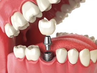
The study evaluated rates of dental implant loss and peri-implantitis as the result of dental implants.

Any dental procedure comes with the chance of infection, but a recent study out of Sweden has discovered that patients with dental implants run a high risk – and that several contributing factors can exacerbate this risk

The research out of the University of Gothenburg aimed to evaluate the correlation between dental implants, implant loss and peri-implantitis, a destructive infection that affects the tissue surrounding dental implants and can result in loss of supporting jawbone. Researchers also explored the relationship between periodontitis and rates of implant loss.

The study consisted of 4,716 randomly selected participants that had all had dental implants in 2003-2004. Researchers sent out a survey and received dental records and charts for 2,765 patients in the study. 596 patients were also examined at a nine-year follow-up appointment. The results found that almost eight percent of patients with dental implants experienced the loss of at least one implant within that timeframe.

More research on dental implants: New discovery can prevent dental implant infections

“Altogether, 7.6 percent of patients had lost at least one implant and 14.5 percent had developed peri-implantitis with pronounced bone loss,” reported Dr. Jan Derks, a researcher at Sahlgrenska Academy. 50 percent of patients presented with some signs of peri-implantitis, but only 14.5% were considered to have moderate to severe implications (equating to a crestal bone loss exceeding 2 mm). The 7.6 percent that had lost an implant showed an average loss of 29 percent of bone support.

Dr. Derks’ research also found that patients with preexisting periodontitis experienced an increased risk of peri-implantitis. Smoking was also identified as a risk factor contributing to early implant loss. The study also reported that “progression of peri-implantitis occurred in a non-linear, accelerating pattern, and, in the majority of cases, the onset of the disease had occurred early.”

More emerging research: Study finds protein can inhibit bone loss from periodontitis

Interestingly, the rate of implant failure did not differ between the general practice and specialty practices. “22% of all patients in the present sample received their implants in a general practice setting, and implant los in this subgroup was not different from outcomes in patients treated in specialist clinics,” the study stated.

“Peri-implantitis appears to develop within a few years and then progresses quickly at an accelerating pace,” said Dr. Derks. He hopes that the information gained from the study can help dentists minimize the risk of peri-implantitis and implant loss.

Dental implants Instructions

Dental implants have been accepted as a viable treatment option for completely and partially edentulous patients.1,2,3 Further improvements toward the successful osseointegration of dental implants have involved modifications to both surface topography and surface chemistry.4 Implant design (i.e., types and dimensions), surgical procedure, implant placement time, and time prior to loading have been shown to influence implant survival rates.5,6,7,8.

Why to choose this treatment dental?

Therefore, risk factors associated with implant failure have become a frequently discussed topic in recent dental research. Among patient factors, male gender, smoking, autoimmune disease, and penicillin allergy have been found to trend toward higher failure rates.9,10 In contrast, short implants in mandibular posterior sites have been found to have a survival rate of 100%, while the same was not true for implants in the maxillary posterior position, wherein 6-mm implants had a survival rate of only 87%.

Most survival study designs are longitudinal and retrospective with many years of follow-up. Their statistical methods use implant failure as the unit of analysis, without considering multiple implant failures in the same patient. Outcomes related to implants in a single patient must be more closely correlated than those in separate patients; ignoring these correlations could result in a bias in p-value computations.11,12,13

Therefore, in this retrospective study, we aimed to evaluate dental implant outcomes and to identify risk factors associated with implant failure over 12 years via dental records of patients attending an educational institution.

Reasons to Upgrade to a Dental X-ray Machine(一)

In most cases digital x-ray machines require less radiation to capture the same image than a traditional film x-ray unit. Digital portable dental x rays machine also has a drastically reduced exposure time in comparison and do not require as many scans to capture the necessary view. This means patients are exposed to less radiation and a more pleasant office visit experience. You’ll end up with shorter wait times and happier patients. With more advanced digital imaging technology, scan times continue to shrink, and you can see almost immediately if you captured what you need to see.

portable dental x rays machine

Digital x-rays are much more eco-friendly

We can’t forget the burden the chemicals needed to develop x-ray film place on the environment. Imagine all those harmful chemicals lying around. What happens to them after you dispose of them? No matter “how safe” you are told the disposal process is, they don’t just go away, nor does their impact on the environment.

Film portable dental x ray unit cost you valuable time.

Film radiography is an entire process alongside the actual scan. There are specific steps for film to be developed. With digital radiography all of these steps can be done away with, freeing you and your staff up to see more patients. Think of it this way. It takes around 10-18 minutes for a traditional portable dental x ray unit to be produced, from initial scan to development. Let’s pretend you average 8 patients a day, and assume all images come out perfectly every time with no need for rescan. That means in total, your practice has spent anywhere from 1.5-2 hours in simply developing and production of x-ray films. That’s potentially 1-2 more patients that your practice could see! Of course this also does not include the time it takes to compute these images.

X Ray Film Viewer Medical Diagnostic E.N.T. Imaging LED Illuminator View Box NEW

Better Image Quality and Manipulation make for better diagnoses

Analog x-ray imaging systems are workhorses that continue to produce quality images. However, just as camera lenses have advanced rapidly in the past decade (think about the quality of your camera phone!) so too have digital x-ray sensors. They continue to become sensitive enough to reduce exposure times, while still capturing an image that can be enhanced and altered to best display exactly the information you need to diagnose your patient. Whereas for film, though their quality is still perfectly adequate, there’s no way to enhance images. It can take several scans before a doctor can confidently and accurately make a buy dental equipment online plan based on traditional analog film x-rays. And as we’ve already pointed out above, rescanning with a film x-ray is much more of a hassle than a digital x-ray.

Making an adjustment and rescanning is simple and hassle free. Rather than sending patients away while x-rays develop, dentists can almost instantly see their digital images, cutting down on the time your patients impatiently sit around in the office. The convenience of capturing another image right then and there results in much happier and satisfied patients, eliminates any potential guesswork, and rapidly improves practice efficiency by streamlining the entire portable dental x rays machine process.


Benefits of Digital Dental X-Rays Used in Dental Treatment

The film is immediately processed and available to view, whereas film takes time to be developed. Less radiation needed to produce the same quality image as film (digital X-rays gives 70% less exposure to radiation than conventional X-rays).

You can enhance the digital image (such as alter brightness and contrast) with a series of processing techniques:

  1. Digital archiving—the ability to store images on a computer
  2. Digital radiography produces larger photos to better source hard-to-see cavities
  3. Digital radiography, though expensive to buy initially, is cheaper and more environmentally friendly in the long run

Dental Portable Digital X-Ray Machine

Grey-scale of digital dental x rays unit for sale offer 256 shades of grey versus 16-25 shades in conventional radiography. Particular benefits of digital radiography for the dentist’s office include improved comfort of intra-oral (photos taken inside the mouth) X-rays, performed by intra-oral dental assistants, and less cranial exposure to radiation. Digital images are also easily stored on a computer and can be transferred to different dentists or orthodontists for referrals. Despite this, there are still certain benefits offered by film radiography, which is why so many dentist offices maintain their older machines.

Benefits of Film Radiography

  1. Offers excellent picture quality
  2. Most dentists’ office are still equipped with conventional radiography equipment, which is perfectly usable and saves money to not change
  3. Film sensors are less thick and rigid than digital sensors, thus offering the patient better comfort when the X-ray sensors go into their mouths
  4. There is no extra training required to use conventional X-ray machines, as it is standard in most dental offices.
  5. Unlike digital X-ray sensors which must use plastic disinfectant covers, conventional film sensors can withstand heat sterilization

X Ray Film LED Illuminator View

Film X-ray photos cannot be altered, whereas digital ones can. Possibility of alteration exposes digital radiography to fraudulent use and legal issues. Digital radiography is still slowly being adapted by dentist offices, but a greater change may soon be coming, with much dental hygienist training teaching the digital process.our dentist equipment for sale ensures that images will be readily available, allowing professionals to streamline the process. The Scan exam provides professionals with the intraoral diagnostic images that they require in seconds – an unprecedented level of speed and clarity. Image settings are predetermined to allow for a smooth and facile process but also remain adjustable to accommodate customized setting preferences. To save you time, money and provide continuity, our imaging plates are designed for repeated use, guaranteeing superior image quality every time.

You Want to Know Something About Dental X-Rays

There are two main types of dental x-rays: intraoral (meaning the x-ray film is inside the mouth) and extraoral (meaning the x-ray film is outside the mouth). Intraoral x-rays are the most common type of dental x-ray taken. These x-rays provide a lot of detail and allow your dentist to find cavities, check the health of the tooth root and bone surrounding the tooth, check the status of developing teeth, and monitor the general health of your teeth and jawbone. Extraoral x-rays show teeth, but their main focus is the jaw and skull. A new set of portable dental x rays machine may be needed to help your dentist detect any new cavities, determine the status of your gum health or evaluate the growth and development of your teeth.

Colorful Wireless Handheld Dental Intra-oral Portable X-Ray Unit BLX-10

If a previous dentist has any radiographs of you, your new dentist may ask you for copies of them. Ask both dentists to help you with forwarding your x-rays. These x-rays do not provide the detail found with intraoral x-rays and therefore are not used for detecting cavities or for identifying problems with individual teeth. Instead, extraoral x-rays are used to look for impacted teeth, monitor growth and development of the jaws in relation to the teeth, and to identify potential problems between teeth and jaws and the temporomandibular joint (TMJ, see temporomandibular disorders for more information) or other bones of the face. If you are a new patient, the dentist may recommend x-rays to determine the present status of your oral health and have a baseline to help identify changes that may occur later. The high cost of x-ray units makes them economically out of reach for some practices, particularly those in rural areas with small patient bases or in developing countries.

HISHINE® Luna Dental X-ray Film Reader LED Specific Wall Desk Mounting

Fortunately, portable radiography systems can bring these services to practices that lack them. But do portable units perform as well as their stationary counterparts? The industry got shaken up a few years ago with the appearance of ”budget” models on various web shops. These machines were offered on prices even 10x lower than the standard brands. Studies in US and UK showed that these products’s radiation emission were way over limits, so additional legislation was passed on the import of portable x-ray machines. Dental x-rays are a useful dentist equipment for sale tool when helping your dentist detect damage and disease not visible during a regular dental exam. How often X-rays should be taken depends on your present oral health, your age, your risk for disease, and any signs and symptoms of oral disease. For example, children may require x-rays more often than adults because their teeth and jaws are still developing and their teeth are more likely to be affected by tooth decay than those of adults. Your dentist will review your history, examine your mouth and then decide whether or not you need x-rays.

Dental Implants – Will They Do the Work?

The niche of dentistry along with the technology that is put to use by the dentists is developing at an alarming pace. There was a time when the commoners had to opt for removable dentures, if they lost their teeth. Times are changing and now we have access to dental implants. What is dental implant motor? What are the various kinds of dental implants? Can I opt for this procedure if I lose a couple of teeth? The queries posed to cosmetic dentists in the real world as well as in the virtual world (internet discussion forums) are also increasing. If you are in need of a dental-implant, the first thing you must be doing is to fix an appointment with the local dentist. Sometimes, expert cosmetic dentists can rectify minor problems with the use of ulterior technology.

2017 New Dental Implant Surgical Brushless Drill Motor Reduction Handpiece

A dentist will examine the patient thoroughly. He will also employ advanced technologies that will generate portable dental x rays machine images of the jawbone and the gums. All these are required to determine the type of dental implant, which will work out to be highly feasible for the patient. Yes, the ‘one size fits all’ paradigm does not work out over here. Many patients would shun away from dental-implant procedures because of the pains. However, you must realize that you are in safe hands. An expert dentist will employ the least painful procedures. I am not stating that the procedure is painless. In the words of expert dentists, dental implants might cause ‘minor discomforts’. Once the process is completed, the dentist might ask the patient to seek respite on painkiller prescriptions. Do not worry, the dentist will provide the same. The healing period varies greatly – if you follow the guidelines cited by the dentist, it will take a maximum of three to four weeks for the pain to subside. I understand that many readers might have various notions when it comes to the success rates of dental implants.

Dental Handheld Portable Digital Green X-Ray Machine System 60W 30 KHz High End

You must shed the belief that this is a new technology. Dental-implantation procedures have been in existence since some years. Throughout these years, dentists have perfected the niche. Hence, the success rate of dental implant procedure is 90 to 95 percent. Does that make you feel better? I do realize that you might have numerous queries surfacing in your mind right now. The best person, who can give you satisfactory answers, is none other than your dentist. It is important to find the right kind of dentist who is well versed with dental implants. Someone who utilizes the latest techniques will be your best bet. The internet is a good place to initiate the search for the finest cosmetic dentists. You must also consider the number of sittings (appointments) with the dentist.

Outfit Your Practice with the Best Portable Dental Unit with Tip

Opening, renovating, or updating your professional space can often be challenging. When you have a medical or dental practice that requires specialized tools and equipment, this is particularly so. When you are looking for a new type of dental unit or other equipment for your practice, you will typically find that you have quite a few different options in designs and features. You will find quality items that perform in the ways that you want, sometimes finding everything that you need in the same place, when you research the different equipment types and the manufacturer.

The first step to choosing dental delivery systems is whether you want a chair or base mounted portable dental delivery units. Chair or base mounted units can be rear, side, ellipse or over the patient. Typical dental delivery systems have automatic activation for at least three handpieces, self-contained waterline with asepsis tubing and purge system, airflow control and a control panel with wet/dry foot control. Some dental delivery systems can integrate various accessories like fiber-optic systems, intraoral cameras, x-ray viewers, lighting and touchpads. There are numerous products to choose from, be sure your vendor offers an extensive warranty, is available for any questions during implementation and offers technical support.

2017 New Greeloy®Portable Dental Mobile Delivery Unit +Air Compressor+LED Fiber HP Tube 6H New

Look into the Manufacturers

Not all manufacturer’s and distributor’s products are designed the same way, and some offer you more options and stability than others. Good things to look for when researching manufacturers is how long they have been in business and how many positive reviews they have. This is essential because if you have a warranty on an item that breaks, you’ll be able to have it fixed or replaced if the company you got the warranty from is still in business. You can also get a good indication of how well its equipment performs and how easy it’s going to be to get replacement parts if needed if you examine the stability of a manufacturer. You will also want to investigate the physical location of the equipment when you go to order it to see how difficult it will be to get it shipped to your location and installed. Much of this information you can find on the company’s website, but you can also find third-party reviews and get advice from your professional contacts. Going with a company with a reputation for quality and sterile manufacturing processes can help you guarantee the safety of your patients as well as the longevity of your equipment.

Finding the  best portable dental unit and other equipment for your practice can be as simple as doing a bit of research into how the various types can help fit your needs. To find the items and features that you are looking for, you can look through online catalogues; you may even be able to get all the items that you need from the same place. You can also find information regarding the manufacturer and distributor online to help you make a knowledgeable decision that will work for you far into the future. Looking for such things as company stability and reputation for quality can help keep your costs low while ensuring that you are providing exceptional service.

How to Use the Dental Surgical Light for Dental Treatment Procedures

In both dental and surgical applications it’s common to find medical professionals utilizing attachable light sources on their loupes. The right amount of loupe light can be an essential component to any medical application. With a clear view, a doctor can perform procedures and exams thoroughly and safely. An attachable clip-on light typically generates light from a source that can be easily clipped-on or stored nearby. Some surgical models are designed in that the cord from the light source runs from a tabletop or wall-mounted platform. And here, we first need to introduce the dental surgical light which is a kind of dental supply for sale.

As any clinician will know, the correct lighting is essential if one is to perform precision-oriented tasks such as dental procedures. During the course of a working day, most dental practitioners are constantly having to adjust their vision to cope with the contrasting light conditions produced by operating lamp and consulting room overhead lighting. To complicate matters further, our eyesight tends to deteriorate with age, meaning that as we age, our eyes require more and more light in to order to perform at the same level. Eyestrain due to poor lighting can hasten optical deterioration as well as lead to eye strain, leading to headaches, fatigue and, if forced to continuously move towards a single light source, neck and back strain. It is therefore critical to ensure that your consulting rooms have dental lighting that minimises the risk of strain whilst also ensuring an optimal view of the patient’s mouth.

Both designs will work, it really just depends on the type of procedure and how your medical environment is set up. It’s important to keep in mind that a good light source can be essential during your medical application. Without proper dental surgical light, medical professionals can sometimes make an oversight when scrutinizing a patient. Loupes, in general, help determine a better diagnosis for patients and loupe light is an additional benefit during these applications. For many dentists, an attachable loupe light is a crucial component in performing precision dentistry. Dentists and dental assistants can often block the overhead light when examining a patient. Loupe lights allow dentists to focus light onto a particular spot in the patient’s mouth, without shining an uncomfortable bright light into the patient’s eyes. The benefits of using an added light source includes the ability to clearly see the precise area of the patient. With this clear visibility, doctor’s can use best practices as they keep their patient safe and provide excellent patient care. If you have any questions or comments regarding loupe light, please let us know in the comment box below!

The right dental lighting should strike a balance between providing enough high intensity illumination to enhance work precision, especially when it comes to things like colour matching for restorations and cosmetic work, whilst minimising glare and other visual irritants caused by phenomena such as strobing and flickering. Ideally, the ratio of contrast between the lighting used to illuminate the treatment site and the area surrounding the patient should never be greater than 1:5 in terms of strength. By way of example, then, if the working area around the patient’s mouth is in the region of 20,000 lux then the lighting in the environment immediately surrounding this should be no less than 4,000 lux. Specialist overhead lighting systems can help to soften this difference in lighting strength whilst simultaneously minimising shadows, glare and flickering and reduce heat emissions to ensure comfort and accuracy.

Information You Should Know About The Endodontic Motors

Endodontic instrumentation has advanced significantly in recent years with a range of powered rotary and reciprocating systems that can make root canal treatment easier to perform.

Endodontic electric motors are a part of this advancement as they provide clinicians with the ability to use special endodontic Nickel-titanium (NiTi) files.

NiTi rotary instrumentation should always be performed with slow-speed, low-torque electric motors with constant and precise torque suited to the canal being treated. Endo motors offer programmable settings, automatic reverse functions designed to reduce file breakage, and even integrated apex locators.

Dental equipments have three times more elastic flexibility in bending and torsion as well as superior resistance to torsional fracture when compared with size stainless-steel files that is why nickel-titanium files gradually force out hand stainless files.

NiTi files have many practical benefits:


— Predictable results every time even when dealing with  canals.

— Time Saving: with practice a standard molar can be totally cleaned and shaped in far less time than hand instrumentation.

— Less post-op pain owing to debris being extruded from the canal during the crown-down technique instead of being pushed through the apex during step-back.

— Less Fatigue for the operator compared to dental equipment.

— Less Transportation of canals.

Teeth with severely curved or S-shape roots should not be instrumented by rotary files to the working length because a NiTi file in a curved canal will have the tendency to straighten itself, resulting in transportation, zipping, and stripping.

Radius of curvature is the most significant factor in terms of predicting cyclic fatigue failure (separation) of the instrument.In other words, the more curved the canal, the greater the risk for separation.

Source from:

Information You Should Know About The Endodontic Motors


What Is The Dental Surgical Light ?

The requirements laid down in national and international standards and draft standards provide helpful guidelines for optimum illumination (Fig. 1, Table I). General room illumination with 500 lx and illumination of the working area with 1000 lx are best achieved with a larger number of fluorescent lamps on the ceiling above and in front of the dental chair. Daylight white lamps with good color rendering are a good combination with changing daylight and the color of light of the operating light. The color of external skin, mucous membrane and teeth appears natural. The eight surgical lights examined differ in quality (Figs. 2-9). The maximum illuminance is between 9000 and 21 000 lx and is thus sufficiently high. The evenness of dental surgical light distribution within an ellipse 9 cm and 18 cm in diameter is between 1 : 4 and 1 : 15 (Figs. 10 and 11). Illuminance can be adjusted to the work in hand by means of controls. If the patient looks into the operating lights (Figs. 2c and 9c), maximum luminances of 5 cd/cm2-20 cd/cm2 occur 8 cm above the illuminance maximum in six operating lights. Luminances of more than 20 cd/cm2 cause squinting and running eyes. A light fitting with more than 200 cd/cm2 should not be used.

Besides the dental surgical light, we can also see the dental examination light during teeth treatment. A sharp fall in illuminance (distinct light/dark threshold) and low luminances to the patient’s eyes can be achieved with very directed light. Very directed light leads to very heavy shadows. Similarly, less specifically directed light leads to softer shadows so that objects in the oral cavity can be discerned easily, but the patient is no longer dazzled. The following operating lights can be recommended if the patient is to suffer as little glare as possible: Den-Tel-Ez Daray, and Belmont Type 040, Faro Sunlight S 70, Ritter Super Starlite; as well as: Chirana Fax, Siemens Sirolux. The following can be recommended for good illumination of the oral cavity: Belmont Type 040, Chirana Fax, Emda Top Spot, Faro Sunlight S 70, Pelton and Crane Light Fantastic Plus, Ritter Super Starlite, Siemens Sirolux. The color temperature, heat radiation, easy handling, stability and price are also important for qualitative assessment.